 ImageGear MDby Accusoft
AccuSoft® ImageGear® MD is a powerful medical imaging toolkit, for both Windows and Linux platforms, that developers use to quickly design applications to read, write, display and transform DICOM (Digital Imaging and Communications in Medicine) medical files while adhering to medical imaging standards.
Medical Formats
The format support of the ImageGear MD component includes loading and saving monochrome, palletized, and true color medical images using the following file formats:
- DICOM 3.0 Part 10-compliant images
- DICOM 3.0 Raw Format (non-Part 10-compliant)
In addition, your application will also support all of the ImageGear supported formats — allowing you to convert an image of a different format to a medical image format, and vice versa.
Read and Write
When reading and writing DICOM image files, ImageGear MD provides the following capabilities:
- Converts to and from other ImageGear supported formats
- Scans 9- to 16-bit grayscale images using TWAIN
- Supports monochrome, grayscale, palletized and true color DICOM images
- Reads and writes both Big Endian and Little Endian byte orders
- Auto-detects DICOM images with or without a Meta-info (Part 10) header
- Reads many out-of-spec images
- Reads 1-bit DICOM files
- Reads and writes multi-frame and Cine files
- Supports both Implicit VR and Explicit VR.
- Reads files with or without group lengths
- Loads and saves DICOM images without data element (DE) loss
- Preserves 16-bit grayscale pixels
- Reads RGB planar images
- Reads and writes JPEG lossy, JPEG lossless, RLE and JPEG 2000 compressed DICOM images
 Display Display
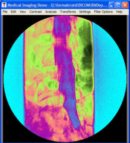
When displaying DICOM image files, ImageGear MD provides the following capabilities:
- Real-time contrast adjustment using Window level, rescale, and gamma correction transforms
- Supports bit depths of 8- to 16-bits per pixel
- Offers pseudo-coloring of the entire image using either pre-defined pseudo-color schemes or your own customized LUTs
- Allows pseudo-coloring of only those pixel values that are over the high threshold, or under the low threshold (allows you to set the thresholds)
- Support for cine loop display of image sequences
Presentation State Objects
ImageGear includes support for Presentation State objects. These objects serve the following purposes within a DICOM file:
- Consistent display of images on various devices and media
- Storage of specific settings for display
- Storage of annotations and additional tags such as Patient/Study/Series information
Presentation State files do not contain images, but rather reference other images for which they provide the above mentioned attributes.
ImageGear provides full support for the Presentation LUT as well as the Grayscale Standard Display Function (GSDF. This allows consistent image display and contrast settings. Additionally, the Presentation State data set can be loaded and merged with the image data set.
Presentation State annotations are also supported. These include:
- Textbox
- Point
- Poly-line
- Spline
- Circle
- Ellipse
These annotations are converted into ImageGear ART objects during reading, and exported back into presentation state data during saving. Graphical objects can be filled or not. Additionally, annotations can be grouped into layers.
Image Processing
When processing medical image files, ImageGear MD provides the following capabilities:
- Converts 16-bit images to 8-bit images and vice versa
- Enables contrast adjustment
- Tabulates up to 16-bit histogram data
- Converts signed to unsigned pixel data
- Swaps bytes of 16-bit pixels
- Supports convolve matrix, sharpen, smooth, edge maps, rotate, resize, flip and crop functions for 16-bit images
- Supports 16-bit images in many baseline ImageGear Image Processing API functions
- Enables shifting up or down of the High bit
- Returns the minimum and maximum pixel values from a chosen area of an image
Data Set
When working with the data set, ImageGear MD provides the following capabilities:
- Complete API for data set creation and removal
- Complete data set access, including the modification, addition and removal of data elements
- Complete support of SQ and multiple levels
- Enables customer-defined tags to be added to the data dictionary
Annotations
The AccuSoft Redlining Toolkit (ART) component, which is included with ImageGear Professional makes an excellent companion to ImageGear MD. It provides the ability to add annotations to a medical image, adding many benefits for end users:
- Add arrows
- Highlight the areas of interest on a medical image
- Place notes or observations on to an image
- Block out (redact) sensitive areas
- And much more
Additionally, annotations can be merged with an image, or kept in a separate file and overlaid on the image at display time. In this way, the original image is never directly altered.
|